Category Archives: Urology Services

appendix surgery in Laxmi Nagar, East Delhi

Appendix Surgery

Appendix Surgery in Laxmi Nagar, East Delhi: Expert Care for Appendicitis

Introduction

Appendix surgery, often performed to treat appendicitis, is a common medical procedure. If you’re in Laxmi Nagar, East Delhi, and require appendix surgery or want to learn more about this procedure, you’ve come to the right place. In this comprehensive guide, we’ll cover the essentials of appendix surgery, including its causes, symptoms, surgical techniques, and where to find expert care.

Understanding Appendicitis

Appendicitis is the inflammation of the appendix, a small, tube-like organ located in the lower right abdomen. When the appendix becomes blocked or infected, it can lead to severe pain and complications.

Signs and Symptoms

  1. Abdominal Pain: The primary symptom of appendicitis is sharp, intense pain in the lower right abdomen.
  2. Loss of Appetite: People with appendicitis often lose their appetite and may feel nauseous.
  3. Fever: A low-grade fever may accompany appendicitis.
  4. Vomiting: Some individuals with appendicitis may experience vomiting.

When to Seek Medical Attention

Appendicitis requires prompt medical attention. If you experience severe abdominal pain, especially in the lower right area, along with other symptoms like fever and vomiting, consult a healthcare professional immediately.

Appendix Surgery

Surgical Procedure

Appendix surgery, also known as appendectomy, involves the removal of the inflamed appendix. There are two primary approaches:

  • Open Appendectomy: This traditional approach involves a single incision in the lower right abdomen to access and remove the appendix.
  • Laparoscopic Appendectomy: This minimally invasive approach uses small incisions and a camera to guide the surgeon in removing the appendix.

Your surgeon will determine the most appropriate technique based on your condition.

Recovery

Recovery from appendix surgery typically involves a short hospital stay and a few weeks of rest. You’ll receive instructions on pain management, dietary guidelines, and when you can resume normal activities.

Frequently Asked Questions (FAQs)

Is appendectomy the only treatment for appendicitis?

Yes, surgical removal of the inflamed appendix is the standard and most effective treatment for appendicitis.

How long does an appendix surgery take?

The duration of the surgery depends on the chosen technique and individual factors but typically ranges from 30 minutes to an hour.

Can I live a normal life without my appendix?

Yes, the appendix is not essential for survival, and most people lead entirely normal lives after its removal.

Are there any complications associated with appendix surgery?

Like any surgery, there are potential risks and complications, but they are relatively rare. Your surgeon will discuss these with you before the procedure.

Where can I find expert appendix surgery in Laxmi Nagar, East Delhi?

For skilled appendix surgery and comprehensive care, consider [Hospital/Clinic Name] in Laxmi Nagar. Our experienced surgical team is dedicated to your well-being.

Conclusion

Appendix surgery in Laxmi Nagar, East Delhi, offers a solution for those suffering from appendicitis. Prompt medical attention and expert surgical care are vital for a successful outcome. If you or a loved one experience symptoms of appendicitis, do not hesitate to seek immediate medical assistance. Trust in the expertise of healthcare professionals to ensure your health and well-being.

Fibroadenoma Treatment in Laxmi Nagar, East Delhi

Fibroadenoma Treatment

Fibroadenoma Treatment in Laxmi Nagar, East Delhi: Your Path to Wellness

Introduction

Fibroadenomas, though typically benign, can cause anxiety and discomfort. If you’re in Laxmi Nagar, East Delhi, and seeking fibroadenoma treatment, you’re on the right path to prioritize your health. This article serves as your guide to fibroadenoma treatment in Laxmi Nagar, covering everything from understanding the condition to available treatment options and where to find compassionate care.

Understanding Fibroadenomas

Fibroadenomas are noncancerous breast lumps that commonly affect women, particularly in their twenties and thirties. While these growths are generally harmless, they can be concerning due to their presence.

Signs and Symptoms

  1. Breast Lump: The most common sign is a palpable lump in the breast that is firm and well-defined.
  2. Smooth Texture: Fibroadenomas often have smooth edges and a rubbery texture.
  3. Painless: Typically, fibroadenomas do not cause pain, but some individuals may experience tenderness or discomfort.
  4. Stable Size: In most cases, these lumps remain stable in size, although they can grow slowly over time.

When to Seek Medical Attention

While fibroadenomas are generally harmless, certain situations warrant medical evaluation:

  • New Lump: If you discover a new breast lump or notice changes in an existing lump, it’s crucial to consult a healthcare professional.
  • Pain or Discomfort: If a fibroadenoma becomes painful or causes discomfort, it’s essential to have it evaluated.
  • Uncertainty or Concern: Whenever you have doubts or concerns about changes in your breast tissue, seek prompt medical advice.

Diagnosis and Treatment

Medical Assessment

A thorough medical assessment is essential for diagnosing fibroadenomas. This may include a physical examination, mammogram, ultrasound, or biopsy.

Treatment Options

Treatment for fibroadenomas varies based on factors such as size, growth, and symptoms. Common treatment options include:

  • Monitoring: Small, stable fibroadenomas may not require immediate treatment but should be monitored regularly by a healthcare professional.
  • Fine Needle Aspiration (FNA): In some cases, doctors use FNA to remove fluid from the lump for diagnostic purposes.
  • Surgical Removal (Excision): Larger or symptomatic fibroadenomas may be surgically removed. Surgical excision can provide relief from discomfort and address cosmetic concerns.

Supportive Care

A fibroadenoma diagnosis can be emotionally challenging. Seek support from healthcare providers who specialize in breast health. They can offer guidance, answer questions, and provide reassurance throughout your journey.

Frequently Asked Questions (FAQs)

Are fibroadenomas cancerous?

No, fibroadenomas are benign (noncancerous) breast tumors.

Can fibroadenomas turn into cancer?

While fibroadenomas themselves are not cancerous, they do not directly transform into cancer. However, they may be associated with a slightly increased risk of breast cancer.

Is surgery the only treatment option for fibroadenomas?

No, surgery is not always necessary. Treatment decisions are made based on individual factors, and monitoring or less invasive procedures may be suitable for some cases.

Will fibroadenoma treatment affect my ability to breastfeed?

In most cases, fibroadenoma treatment should not impact your ability to breastfeed. Discuss any concerns with your healthcare provider.

Where can I find specialized care for fibroadenoma treatment in Laxmi Nagar, East Delhi?

For expert diagnosis, treatment, and compassionate support for fibroadenoma in Laxmi Nagar, consider [Hospital/Clinic Name]. Our dedicated team is committed to providing comprehensive care tailored to your specific needs.

Conclusion

Fibroadenoma treatment in Laxmi Nagar, East Delhi, ensures you receive the care and support needed for your breast health. Remember that early diagnosis and appropriate medical attention can offer peace of mind and promote your overall well-being. If you suspect a fibroadenoma or have concerns about your breast health, reach out to experienced healthcare professionals who can guide you through diagnosis, treatment, and support.

hernia surgery in East Delhi

Hernia Surgery

Hernia Surgery in East Delhi: Your Path to a Pain-Free Life

Introduction

Hernias can be painful and disruptive to your daily life. Fortunately, if you’re in East Delhi, you have access to exceptional hernia surgery options. In this article, we’ll explore everything you need to know about hernia surgery in East Delhi, from understanding the condition to the recovery process. Trust in our expert surgeons to guide you towards a pain-free life.

Understanding Hernias

Before delving into hernia surgery, let’s grasp the basics. A hernia occurs when an organ or tissue pushes through a weak spot in the muscle or connective tissue. Common types of hernias include inguinal (groin) hernias, umbilical (belly button) hernias, and incisional hernias (resulting from previous surgical incisions).

Signs and Symptoms

  1. Visible Bulge: One of the primary signs of a hernia is a noticeable lump or bulge in the affected area.
  2. Pain or Discomfort: Hernias often cause discomfort or pain, especially when lifting heavy objects or straining.
  3. Burning or Aching Sensation: Some individuals experience a burning or aching sensation at the hernia site.

When to Consider Surgery

While not all hernias require immediate surgery, certain factors may indicate the need for intervention. These include:

  • Increasing Pain: If the pain associated with your hernia becomes severe or persistent, it’s time to consult a surgeon.
  • Size and Growth: Larger hernias or those that are increasing in size typically require surgical repair.
  • Obstruction or Strangulation: If a hernia becomes incarcerated (trapped) or strangulated (blood supply cut off), it’s a medical emergency necessitating surgery.

The Hernia Surgery Process

Preoperative Assessment

Before your surgery, you’ll undergo a thorough evaluation to assess your overall health and determine the best approach for your hernia repair.

Surgical Techniques

There are different surgical techniques for hernia repair, including open surgery and minimally invasive laparoscopic procedures. Your surgeon will recommend the most suitable option based on your hernia type and medical history.

Recovery and Aftercare

Recovery from hernia surgery varies depending on the surgical technique and individual factors. However, here are some general guidelines:

  • Pain Management: You may experience some discomfort post-surgery, but pain can be managed with medication.
  • Activity Restrictions: Your surgeon will provide specific instructions regarding activities and lifting restrictions during your recovery.
  • Dietary Guidelines: Following surgery, you may need to adjust your diet temporarily to avoid straining the surgical site.

Frequently Asked Questions (FAQs)

How long does hernia surgery take?

The duration of hernia surgery varies but typically ranges from 30 minutes to 2 hours, depending on the complexity.

Is hernia surgery safe?

Yes, hernia surgery is generally safe. Your surgeon will discuss the potential risks and benefits with you before the procedure.

Can hernias come back after surgery?

While recurrence is possible, modern surgical techniques have reduced the risk significantly. Follow your surgeon’s postoperative instructions to minimize this risk.

When can I return to work after hernia surgery?

Your return to work will depend on the type of work you do and your individual healing progress. Most patients can resume light activities within a week or two.

Is hernia surgery covered by insurance?

In many cases, hernia surgery is covered by insurance. It’s advisable to check with your insurance provider for specific details.

Where can I find expert hernia surgeons in East Delhi?

You can find experienced hernia surgeons at [Hospital/Clinic Name] in East Delhi. Schedule a consultation to discuss your hernia and treatment options.

Conclusion

Hernia surgery in East Delhi offers a pathway to a pain-free and healthier life. If you’re experiencing the discomfort and inconvenience of a hernia, consult with our expert surgeons to explore your treatment options. With modern techniques and compassionate care, you can regain your well-being and get back to enjoying life to the fullest.

Abdominal Wall Hernia Surgery

What is abdominal wall hernia surgery?

An abdominal wall hernia occurs when a part of the intestine or fat protrudes through the opening of the abdominal muscles. Wall hernias can be located anywhere in the abdomen, although the most frequent locations are: the navel, the inguinal region or at the site of a previous incision for abdominal surgery. The main complication of the hernia is that its content is trapped, compromising the blood supply and requiring urgent surgery.

Abdominal wall surgery is a common surgery and carries a small risk of complications. In most cases, you will be able to go home the same day or the next morning; except for large hernias. With this surgery it is possible to repair the opening or muscular defect, using in most cases a prosthesis that gives consistency to the tissue.

Previous considerations

Before any surgery, an assessment should be carried out by the anesthetist where they will advise you which medication you should stop taking and which you should continue taking. Sometimes you must be evaluated by another specialist if you have underlying diseases.

In the case of anticoagulant treatment or treatment that facilitates bleeding, the anesthesiologist, haematologist, cardiologist or family doctor will inform about the attitude to follow.

In the case of smoking, the habit should be stopped because it facilitates anesthetic management, smoker patients have more complications than smokers in anesthetic procedures.

It is recommended to perform moderate-intensity exercise before any intervention, unless there is a specific contraindication, such as walking at least 60 minutes a day.

How is the preparation prior to the intervention?

FOOD AND MEDICINE

In preparation for abdominal wall hernia surgery, your surgeon may ask you to:

  • Do not eat anything before surgery. You may have a sip of water with your medications, but avoid eating and drinking for at least six hours before surgery.
  • Discontinue certain medications and supplements. Talk to your doctor about all medications and supplements you take. You continue to take most medications as directed by your doctor. Your doctor may ask you to stop certain medications and supplements because they can increase your risk of bleeding.

How is the surgery performed? (type of incision, resection, type of drainage, anesthesia)

BEFORE THE INTERVENTION

Abdominal hernia surgery can be performed under general, spinal, or local anesthesia. Numbing medications will be given intravenously in your arm or through a catheter in your spine. Once the medications take effect, the health care team will insert a tube down your throat to help you breathe if your case ultimately benefits from a general anesthetic. Your surgeon will perform the surgery using a laparoscopic or open procedure.

DURING THE PROCEDURE

Depending on your situation, your surgeon will recommend one of two surgical approaches:

Minimally invasive surgery (laparoscopic hernioplasty)

During laparoscopic surgery, the surgeon makes three or four small incisions in the abdomen. A tube with a tiny video camera is inserted into your abdomen through one of the incisions. The surgeon watches a video monitor in the operating room while using surgical tools inserted through the other incisions in the abdomen to repair your muscle defect using a prosthesis (mesh). This intervention can take one or two hours.

A laparoscopic hernioplasty is not appropriate for everyone. In some cases, the surgeon may start with a laparoscopic approach and determine that a larger incision is necessary due to scar tissue from operations, previous complications, or an inability to safely continue with the procedure.

Traditional surgery (open hernioplasty)

During open abdominal hernia surgery, the surgeon makes an incision over the hernia itself. The muscle and tissue are retracted to reveal the defect, which will then be repaired with the use of a prosthesis (mesh).

The incision is sutured and you are taken to a recovery area. An open hernioplasty takes one to two hours. It is usually performed when the procedure cannot be carried out with maximum safety through the laparoscopic approach or due to the characteristics of the hernia.

Sometimes it is necessary to place a tube or drain with an outlet through the skin to allow the discharge of fluid from the area where the hernia was. This tube will be removed prior to discharge home.

What happens after the surgery?

After surgery, the patient slowly awakens from the effects of the anesthetic drugs, so they may have a feeling of not remembering the process. He will spend a short period of time in an area called post-anesthetic recovery, before being taken to his room.

HOW IS THE RECOVERY IN THE HOSPITAL?

It is possible to feel nausea or abdominal pain after the intervention but they will be controlled with the prescribed medication. After about 6 hours, you will begin with the intake of liquids and then solid foods. We recommend sitting down and starting to walk about 8 hours after the intervention. You will be supervised by nursing staff at all times.

If your surgery was performed in the morning and you have no previous illnesses, you may be discharged at the end of the day. If your surgery took place in the afternoon, you will usually leave the following noon. Hospital admission may last for several days in large hernias.

HOW IS THE RECOVERY AT HOME?

Our premise is to be active in recovery. For this reason, we encourage the patient to walk daily, we recommend walking at least 60 minutes each day both inside and outside the house. Important physical efforts that may condition wound healing and prosthesis fixation (mesh) should always be avoided.

When you are discharged home, you will be prescribed medication to make you feel as comfortable as possible. You will resume your previous medication following the surgeon’s recommendations, since some drug may not be recommended in the first days after surgery. Normally the taking of painkillers is indicated to control the pain and sometimes the injection of heparin to avoid the appearance of thrombi in your legs.

In relation to food, we recommend the first week to avoid copious meals. Subsequently, a usual diet.

You can wet your wounds when you wash yourself and then gently dry them with the application of any antiseptic (chlorhexidine, povidone-iodine, crystalmine…). These wounds should be evaluated by nursing around 7 and 10 days after surgery.

Virtually full recovery can take approximately two to three weeks in laparoscopic hernioplasty. However, in open hernioplasty, once at home, full recovery can take four weeks.

What are the risks of hernioplasty?

The normal thing is that your intervention proceeds without incidents but you must know the potential complications.

Infrequent and infrequent risks: Infection, bleeding or fluid collection in the surgical wound. Phlebitis. Acute urine retention. Hematoma. Prolonged pain in the area of ​​the operation. Laparoscopic surgery may cause gas extension to the subcutaneous tissue or other areas and referred pain, usually to the shoulder.

Infrequent and serious risks: Prolonged postoperative pain due to nerve damage. Mesh rejection. Hernia reproduction. Vascular injury. Due to laparoscopic surgery, there may be vascular injuries, injuries to neighboring organs, gas embolism and pneumothorax.

These complications are usually resolved with medical treatment (medicines, serums, etc.), but they may require a reoperation, usually an emergency, and in exceptional cases death may occur.

Your risk of complications depends on your overall health and the reason you’re having abdominal hernia surgery.

Adrenal Gland Surgery

What is adrenalectomy?

An adrenalectomy is surgery to remove one or both adrenal glands.

There is an adrenal gland resting on each of the kidneys. The two adrenal glands produce various hormones that help regulate metabolism, the immune system, blood pressure, blood glucose, and other essential functions.

Most adrenal tumors are noncancerous (benign). You may need surgery (adrenalectomy) to remove an adrenal gland if the tumor produces excess hormones or is large in size (greater than 4 to 5 centimeters). If you have a cancerous (malignant) tumor, you may also need an adrenalectomy. This malignant tumor can come from the adrenal gland itself or from another organ, such as the kidney or lung.

If both adrenal glands are removed, you will need to take hormonal medications. If only one gland is removed, the remaining gland will take over.

Previous considerations

Before any surgery, an assessment should be carried out by the anesthetist where they will advise you which medication you should stop taking and which you should continue taking. Sometimes you must be evaluated by another specialist if you have underlying diseases.

In the case of anticoagulant treatment or treatment that facilitates bleeding, the anesthesiologist, haematologist, cardiologist or family doctor will inform about the attitude to follow.

In the case of smoking, the habit should be stopped because it facilitates anesthetic management, smoker patients have more complications than smokers in anesthetic procedures.

It is recommended to perform moderate-intensity exercise before any intervention, unless there is a specific contraindication, such as walking at least 60 minutes a day.

Sometimes, after a detailed study by the endocrinologist, you must take a specific medication to control the function of your adrenal glands. This will help better control of your vital functions by the anesthesiologist during surgery.

How is the preparation prior to the intervention?

FOOD AND MEDICINE

In preparation for an adrenalectomy, your surgeon may ask you to do the following:

  • Do not eat anything before surgery. You may have a sip of water with your medications, but avoid eating and drinking for at least six hours before surgery.
  • Discontinue certain medications and supplements. Talk to your doctor about all medications and supplements you take. You continue to take most medications as directed by your doctor. Your doctor may ask you to stop certain medications and supplements because they can increase your risk of bleeding.
  • Take certain drugs. Your doctor may prescribe a specific medication to take a few days before the procedure to control the function of your adrenal glands.

How is the surgery performed? (type of incision, resection, type of drainage, anesthesia).

BEFORE THE INTERVENTION

An adrenalectomy is performed under general anesthesia, so you will not be conscious during the procedure. Numbing medications are given intravenously into the arm. Once the medications take effect, the health care team will insert a tube down your throat to help you breathe. Your surgeon will perform the adrenalectomy using a laparoscopic or open procedure.

DURING THE PROCEDURE

Depending on your situation, your surgeon will recommend one of two surgical approaches:

Minimally invasive (laparoscopic) adrenalectomy

During a laparoscopic adrenalectomy, the surgeon makes four or five small incisions in the abdomen. A tube with a tiny video camera is inserted into your abdomen through one of the incisions. The surgeon watches a video monitor in the operating room while using surgical tools inserted through the other incisions in the abdomen to remove the adrenal gland. The incisions are then sutured and you are transferred to a recovery area and sometimes to an intensive care unit if the indication for surgery is because your adrenal glands produce excess hormones. A laparoscopic adrenalectomy takes one to two hours.

A laparoscopic adrenalectomy is not appropriate for everyone. In some cases, the surgeon may start with a laparoscopic approach and determine that a larger incision is necessary due to scar tissue from operations, previous complications, or an inability to safely continue with the procedure.

Traditional (open) adrenalectomy.

Doctors usually reserve open surgery for large or cancerous (malignant) tumors.

During an open adrenalectomy, the surgeon makes an incision about 15 centimeters in the abdomen below the ribs on the side where the diseased gland is located. Muscle and tissue are retracted to reveal and remove the adrenal gland. On the right side it is located below the liver and on the left, behind the pancreas.

The incision is sutured and you are taken to a recovery area. An open adrenalectomy takes about two hours. It is usually performed when the procedure cannot be carried out with maximum safety through the laparoscopic approach.

Sometimes it is necessary to place a tube or drain inside the abdomen with an outlet through the skin to allow the discharge of fluid from the area where the adrenal gland was. This tube will be removed prior to discharge home.

What happens after the surgery?

After surgery, the patient slowly awakens from the effects of the anesthetic drugs, so they may have a feeling of not remembering the process. He will spend a short period of time in an area called post-anesthetic recovery, before being taken to his room.

HOW IS THE RECOVERY IN THE HOSPITAL?

It is possible to feel nausea or abdominal pain after the intervention but they will be controlled with the prescribed medication. After about 6 hours, you will begin with the intake of liquids and then solid foods. We recommend sitting down and starting to walk about 8-12 hours after the intervention. You will be supervised at all times by nursing staff.

It will usually go away in one or two days after the intervention.

HOW IS THE RECOVERY AT HOME?

Our premise is to be active in recovery. For this reason, we encourage the patient to walk daily, we recommend walking at least 60 minutes each day both inside and outside the house. Important physical efforts that may affect wound healing should always be avoided.

When you are discharged home, you will be prescribed medication to make you feel as comfortable as possible. You will resume your previous medication following the surgeon’s recommendations, since some drug may not be recommended in the first days after surgery. Normally the taking of painkillers is indicated to control the pain and the injection of heparin to avoid the appearance of thrombi in your legs.

In relation to food, we recommend the first week to avoid copious meals. Later you could eat your usual diet before the intervention.

You can wet your wounds when you wash yourself and then gently dry them with the application of any antiseptic (chlorhexidine, povidone-iodine, crystalmine…). These wounds should be evaluated by nursing around 7 and 10 days after surgery.

Virtually full recovery can take approximately two to three weeks for laparoscopic adrenalectomy. However, with open adrenalectomy, once at home, full recovery may take four to six weeks.

What are the risks of adrenalectomy?

The normal thing is that your intervention proceeds without incidents but you must know the potential complications.

Less serious and frequent risks: Infection or bleeding of the surgical wound. Phlebitis. Urinary retention. Transient digestive disturbances. Prolonged pain in the area of ​​the operation. Pleural effusion. Laparoscopic surgery may cause gas extension to the subcutaneous tissue or other areas and referred pain, usually to the shoulder.

Infrequent and serious risks : Bleeding or intra-abdominal infection. Intestinal obstruction. Cardiocirculatory and hydroelectrolytic alterations due to changes in hormonal levels. Disease recurrence. Due to laparoscopic surgery, there may be vascular injuries, injuries to neighboring organs, gas embolism and pneumothorax.

These complications are usually resolved with medical treatment (medicines, serums, etc.), but they may require a reoperation, usually an emergency, and in exceptional cases death may occur.

Your risk of complications depends on your overall health and the reason you’re having an adrenalectomy.

Anal Fissure Surgery

What is anal fissure surgery?

An anal fissure is a small tear in the tissue (mucosa) that lines the anus. Anal fissures often cause pain and bleeding during bowel movements. You may also feel spasms in the ring of muscle at the end of your anus (anal sphincter).

Most people with anal fissure respond to hygienic-dietary measures and pharmacological treatment, but sometimes, if there is a good response to them, they may need surgery.

Previous considerations.

Before any surgery, an assessment should be carried out by the anesthetist where they will advise you which medication you should stop taking and which you should continue taking. Sometimes you must be evaluated by another specialist if you have underlying diseases.

In the case of anticoagulant treatment or treatment that facilitates bleeding, the anesthesiologist, haematologist, cardiologist or general practitioner must inform the attitude to be followed.

In the case of smoking, the habit should be stopped because it facilitates anesthetic management, smoker patients have more complications than smokers in anesthetic procedures.

It is recommended to perform moderate-intensity exercise before any intervention, unless there is a specific contraindication, such as walking at least 60 minutes a day.

How is the preparation prior to the intervention?

FOOD AND MEDICINE

In preparation for anal fissure surgery, your surgeon may ask you to:

  • Do not eat anything before surgery. You may have a sip of water with your medications, but avoid eating and drinking for at least six hours before surgery.
  • Discontinue certain medications and supplements. Talk to your doctor about all medications and supplements you take. You continue to take most medications as directed by your doctor. Your doctor may ask you to stop certain medications and supplements because they can increase your risk of bleeding.

How is the surgery performed? (type of incision, resection, type of drainage, anesthesia)

BEFORE THE INTERVENTION

An anal fissure surgery can be performed under general or spinal anesthesia. Numbing medications will be given intravenously in your arm or through a catheter in your spine. Once the medications take effect, the health care team will insert a tube down your throat to help you breathe if your case ultimately benefits from a general anesthetic. Your surgeon will then perform the surgery.

DURING THE PROCEDURE

Lateral internal sphincterotomy.  This technique consists of making a partial cut of the muscle fibers that form the anal sphincter, which will allow less tension to exist on the anal fissure and achieve its healing.

What happens after the surgery?

After surgery, the patient slowly awakens from the effects of the anesthetic drugs, so they may have a feeling of not remembering the process. He will spend a short period of time in an area called post-anesthetic recovery, before being taken to his room.

HOW IS THE RECOVERY IN THE HOSPITAL?

It is possible to feel nausea or abdominal pain after the intervention but they will be controlled with the prescribed medication. After about 6 hours, you will begin with the intake of liquids and then solid foods. We recommend sitting down and starting to walk about 8 hours after the intervention. You will be supervised at all times by nursing staff.

If your surgery was performed in the morning and you have no previous illnesses, you may be discharged at the end of the day. If your surgery took place in the afternoon, you will usually leave the following noon.

HOW IS THE RECOVERY AT HOME?

When you are discharged home, you will be prescribed medication so that you feel as comfortable as possible, so you will have painkillers for when you have pain. Sometimes you may need heparin injection to prevent the appearance of thrombi in your legs. You will resume your previous medication following the surgeon’s recommendations, since some drug may not be recommended in the first days after surgery.

In relation to food, we recommend a diet rich in fiber (vegetables, salads, fruit…) together with an abundant intake of water to ensure that your stools are of a soft consistency and produce the least anal pain when defecating. You may need fiber or laxatives if you have significant constipation after the intervention.

We will recommend sitz baths with warm water and neutral soap several times a day and after defecation. This will ease your discomfort. Avoid using toilet paper.

Walk every day and as you feel less pain, increase the time you dedicate to it. Avoid physical exertion until the surgeon assesses you.

Virtually full recovery can take approximately two weeks after surgery.

What are the risks of anal fissure surgery?

The normal thing is that your intervention proceeds without incidents but you must know the potential complications.

Less serious and frequent risks: Bleeding and infection of the wound, edema of the anus, water retention of urine, prolonged pain in the area of ​​the operation.

Infrequent and serious risks: Significant infection of the perineum, incontinence to qases and even feces, stenosis of the anus, reproduction of the fissure.

death.and even feces. Anus stenosis. Reproduction of the fistula.

These complications are usually resolved with medical treatment (medications, serums, etc.), but they may require a reoperation, and in exceptional cases death may occur.

Your risk of complications depends on your overall health and the reason you’re having fistula surgery.

Bariatric Surgery

What is bariatric surgery?

Bariatric and metabolic surgery with a set of surgical procedures whose objective is the treatment of obesity and associated diseases through alterations in the gastrointestinal tract, together with changes in lifestyle, allows weight loss that is maintained in the long term. . In addition, the remission of multiple metabolic diseases associated with obesity.

Bariatric and metabolic surgery is, without a doubt, the most efficient and safest option to combat obesity, with long-lasting results. In addition, it has become the best option to eliminate or control diseases associated with morbid obesity such as diabetes mellitus, high blood pressure, and dyslipidemia.

It is the ideal intervention for people who suffer from certain physical limitations as a result of obesity. It is applicable to patients with a Body Mass Index (BMI) around 30 points kg/m2.

It is also the solution for patients suffering from type II diabetes mellitus. With the application of any of the bariatric techniques, the patient not only decreases body measurements, but also changes to a better, healthier lifestyle; and as a result, she will feel and look great.

Types of bariatric surgery

There are various types of bariatric procedures, performed with first-class technology in our institution and with a high level of safety, no procedure is better than another, the ideal is to carry out an evaluation with the specialist in order to clarify all your doubts and choose the procedure. most appropriate to meet the needs of the patient. These are the most common types of interventions:

  • Gastric bypass

One of the most performed procedures that consists of an alteration of the amount of food ingested and its absorption through a change in the disposition of the gastrointestinal tract, considerably reducing the calories ingested.

To do this, the surgeon cuts the upper part of the stomach, sealing the rest. The result is a bag the size of a walnut with the capacity to hold approximately an ounce of food.

Finally, the small intestine is cut and sewn directly into the new stomach. In this way, when food is ingested, it will pass through this new digestive system.

It is a very safe type of surgery with excellent results, with a fairly high effectiveness rate, which is why it is called the gold standard of bariatric techniques. That is, with gastric bypass you will be satisfied with less food; but these will be absorbed to a lesser extent; thereby achieving substantial and accelerated weight loss.

At the Metropolitan Clinic we perform bariatric procedures similar to those applied in advanced countries

  • Gastric sleeve

Also called sleeve gastrectomy, it consists of removing a part of the stomach and leaving only a section in the form of a “long, thin tube”, performed through a stapling system. This surgery reduces the amount of food that the stomach can receive, making the patient feel full with less food.

Removing part of the stomach affects some gastrointestinal hormones, which are involved in the process of hunger and metabolism, reducing the feeling of hunger for a certain time.

The gastric sleeve is only restrictive. That is, it reduces the stomach by about 1/3 of its volumetric dimensions. Its name comes from the fact that the new anatomy given to the organ is similar to a tube. In addition, the area that is removed is the largest producer of ghrelin, the appetite hormone; With the extraction of these cells you will be able to feel full faster and thus achieve your desired weight loss.

  • Adjustable gastric band

In this bariatric surgery, the specialist places a ring with an internal inflatable band in the upper part of the stomach, creating a small bag. As in the previous procedures, the patient feels satiated faster when eating.

This gastric band has a circular balloon inside that is filled with a saline solution, so the surgeon can adjust the size of the bag.

Currently, it is one of the procedures that is performed less frequently compared to the previous ones, since it has been associated with more complications, predominantly the need to remove the band due to the patient’s intolerance to food.

  • Biliopancreatic diversion with duodenal switch

It is also known as “composite surgery” and involves two separate procedures. On the one hand, it is similar to the gastric sleeve and on the other hand, it divides the small intestine into two sections.

In this way, food moves through a section without going through most of the small intestine. This helps reduce the total calories and nutrients absorbed by the body.

The advantage of this intervention for patients is considerably greater weight loss than previous procedures. Even so, it is likely to cause complications in the body by not receiving the vitamins, minerals, and proteins that it needs. This is the reason why it is not done very often.

  • Ellipse gastric balloon

New technique that does not require surgery, endoscopy or anesthesia. The ellipse gastric balloon procedure consists of introducing, from the mouth, a capsule that contains a balloon which will be filled with physiological solution, making you feel satiated by occupying it inside the stomach. Patients spontaneously ingest the capsule guided by the specialist.

The gastric balloon that is inserted into the stomach has a vegan film that makes it biodegradable, so it will only remain in the body for 4 months.

Likewise, when the film that covers it begins to deteriorate, the gastric balloon will break and it will be excreted normally by the digestive system.

The advantage of this method is that it helps maintain a restrictive eating process for 4 months, which gives greater satiety to those who carry it with a smaller amount of food. Like the other procedures, it must be accompanied by the support of nutritionists, physiotherapists and psychologists.

Who is a candidate?

In general, bariatric surgery is the best option if you have any of these conditions:

  • The body mass index (BMI) is 40 or more, resulting in extreme obesity.
  • If your BMI is between 35 and 39.9 and you have a serious weight-related health problem, such as type 2 diabetes, high blood pressure, or sleep apnea.
  • In some cases, you may qualify for surgery if you have a BMI of less than 34.

Every patient should be evaluated to determine the best treatment for their condition.

Some specialists will ask the patient to make some changes, which consist of permanently modifying their lifestyle to a healthier one.

Results of bariatric surgery

All bariatric interventions achieve long-term weight loss. This amount will depend on the type of surgery performed and the changes in the patient’s habits.

In addition, it also helps to solve health problems related to obesity, which includes:

  • Heart disease
  • high blood pressure
  • Obstructive sleep apnea
  • Type 2 diabetes
  • Non-alcoholic fatty liver disease (NAFLD) or non-alcoholic steatohepatitis (NASH)
  • Gastroesophageal reflux disease (GERD)
  • Osteoarthritis (joint pain)

Finally, a person’s ability to perform daily activities that they previously could not due to excess weight is improved, which considerably improves their quality of life. Lose weight, gain health.

Surgery complications

Like any surgical procedure, it has its complications. The risks are almost always related to the severity of the conditions that the patient previously presents. These are the main ones:

  • pulmonary embolism
  • internal bleeding
  • Formation of a fistula
  • Vomiting, diarrhea, bloody stools

Almost always, these complications appear while the patient is hospitalized after the intervention, so the medical staff can solve it immediately. At the Metropolitan Clinic we have the best technology to guarantee low complication rates and effective resolution of any complication.

Likewise, some patients may experience nutritional complications such as anemia, folic acid, calcium and vitamin B12 deficiency, which is why continuous follow-up after surgery is vital.

Bile Duct Surgery

What is bile duct surgery?

The bile ducts connect the liver with the gallbladder and the small intestine. Surgery is carried out to remove the part of the bile duct that contains the tumor, both malignant and benign. Surgery may also be recommended to prevent complications or because of the possibility of malignancy.

Sometimes, in addition to the resection of the bile duct, it must be associated with the removal of a part of the liver.

Redrainage of bile from the liver is done using a loop of intestine sewn into the bile duct that remains in the liver.

Previous considerations.

Before any surgery, an assessment should be carried out by the anesthetist where they will advise you which medication you should stop taking and which you should continue taking. Sometimes you must be evaluated by another specialist if you have underlying diseases.

In the case of anticoagulant treatment or treatment that facilitates bleeding, the anesthesiologist, haematologist, cardiologist or family doctor will inform about the attitude to follow.

Normally you should be evaluated by Hematology before the intervention since they must draw blood to study your blood group and make a reserve of blood bags for the day of the intervention. In this surgery, bleeding is an important complication, so we must offer you maximum safety. For your peace of mind, most of our patients do not require a blood transfusion.

In the case of smoking, the habit should be stopped because it facilitates anesthetic management, smoker patients have more complications than smokers in anesthetic procedures.

It is recommended to perform moderate-intensity exercise before any intervention, unless there is a specific contraindication, such as walking at least 60 minutes a day.

How is the preparation prior to the intervention?

FOOD AND MEDICINE

In preparation for a hepatectomy, your surgeon may ask you to:

  • Do not eat anything before surgery. You may have a sip of water with your medications, but avoid eating and drinking for at least six hours before surgery.
  • Discontinue certain medications and supplements. Talk to your doctor about all medications and supplements you take. You continue to take most medications as directed by your doctor. Your doctor may ask you to stop certain medications and supplements because they can increase your risk of bleeding.

How is the surgery performed? (type of incision, resection, type of drainage, anesthesia)

BEFORE THE INTERVENTION

Bile duct surgery is performed under general anesthesia, so you won’t be conscious during the procedure. Numbing medications are given intravenously into the arm. Once the medications take effect, your health care team will insert a tube down your throat to help you breathe, place a catheter to monitor your urine, and place lines in your neck and wrist to administer drugs and learn about your heart function.

You may have an epidural catheter or injection into your spine, as well as local nerve blocks in your abdominal wall. These procedures allow you to recover with minimal pain and discomfort after surgery and help reduce the amount of opioid pain medication you’ll need.

Your surgeon will perform bile duct surgery using a laparoscopic or open procedure.

DURING THE PROCEDURE

Depending on your situation, your surgeon will recommend one of two surgical approaches:

Bile duct surgery with a minimally invasive approach (laparoscopic)

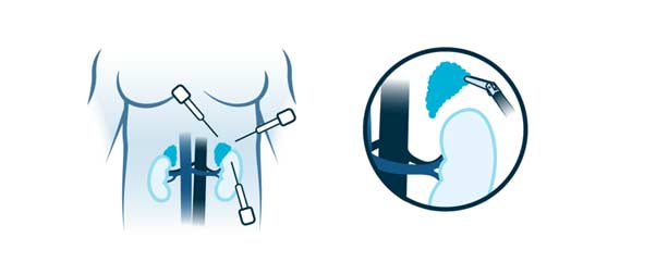
This approach route is used in very selected cases since the technical difficulty is very high. The surgeon makes four to six small incisions in the abdomen. A tube with a tiny video camera is inserted into your abdomen through one of the incisions. The surgeon watches a video monitor in the operating room while using surgical tools inserted through the other incisions in the abdomen to remove the diseased part of the bile duct.

One of the incisions will be enlarged to extract the excised piece. The incisions are then sutured and you are moved to a recovery area. A laparoscopic biliary surgery takes between three and six hours. Very specialized instruments are used to prevent bleeding during the intervention.

In some cases, the surgeon may start with a laparoscopic approach and determine that a larger incision is necessary due to scar tissue from operations, previous complications, or an inability to safely continue with the procedure.

Bile duct surgery with traditional (open) approach.

The surgeon makes an incision about 20 centimeters in the abdomen below the ribs on the right side. Muscle and tissue are retracted to reveal the liver and bile duct. Sometimes, in addition to the resection of the bile duct, it must be associated with the removal of a part of the liver. Redrainage of bile from the liver is done using a loop of intestine sewn into the bile duct that remains in the liver.

The incision is sutured and you are transferred to a post-anesthetic recovery area. An open cholecystectomy takes between three and six hours. Very specialized instruments are used to prevent bleeding during the intervention. It is usually performed when the procedure cannot be carried out with maximum safety through the laparoscopic approach due to the complexity of the case.

Sometimes it is necessary to place a tube or drain inside the abdomen with an outlet through the skin to allow the discharge of fluid from the area where the diseased part of the liver was. This tube will be removed before discharge home, with exceptions.

What happens after the surgery?

After surgery, the patient is transferred to the Intensive Care Unit (ICU) where he will remain for 24 hours if there are no incidents. It is possible that the anesthesiologist will remove the connection to a breathing machine in the operating room or the doctor in charge of your surveillance in the ICU will do it. You will slowly wake up from the effects of the anesthetic drugs so you may have a feeling of not remembering the process. After your stay in this unit you will then be transferred to your room.

HOW IS THE RECOVERY IN THE HOSPITAL?

It is possible to feel nausea or abdominal pain after the intervention but they will be controlled with the prescribed medication. After about 24 hours, he will begin with the intake of liquids and the next day solid foods. We recommend sitting down and starting to walk about 24 hours after the intervention. The catheter that is inserted into your bladder will be removed in 1 or 2 days. At all times you will be supervised and helped by nursing staff.

If your surgery progresses on the floor without any incident, you will remain hospitalized for between 5 and 10 days.

HOW IS THE RECOVERY AT HOME?

Our premise is to be active in recovery. For this reason, we encourage the patient to walk daily, we recommend walking at least 60 minutes each day both inside and outside the house. Important physical efforts that may affect wound healing should always be avoided.

When you are discharged home, you will be prescribed medication to make you feel as comfortable as possible. You will resume your previous medication following the surgeon’s recommendations, since some drug may not be recommended in the first days after surgery. Normally the taking of painkillers is indicated to control the pain and the injection of heparin to avoid the appearance of thrombi in your legs.

In relation to food, we recommend the first week to avoid copious and fat-rich meals. Subsequently, all types of food will be progressively reintroduced, with possible intolerance of some of them, so their intake will be suspended and they will be tried again in the following weeks. Occasionally diarrhea may appear that usually lasts a few days or a week in relation to the new situation of the digestive system.

You can wet your wounds when you wash yourself and then gently dry them with the application of any antiseptic (chlorhexidine, povidone-iodine, crystalmine…). These wounds should be evaluated by nursing around 7 and 10 days after surgery.

Virtually full recovery can take approximately three to four weeks in laparoscopic surgery. However, in open surgery, once at home, full recovery can take four to six weeks. It will depend on your physical condition before surgery and the complexity of the operation.

What are the risks of bile duct surgery?

Bile duct surgery is a technically difficult surgery, even more so if it is associated with the removal of part of the liver, and requires significant experience on the part of the surgeon. Extensive research shows that surgeries result in fewer complications when performed by highly experienced surgeons at centers that perform many of these operations.

Infrequent and frequent risks : Infection or bleeding of the surgical wound, acute urinary retention, phlebitis, intestinal rhythm disorders. Prolonged pain in the area of ​​the operation.

Infrequent and serious risks: Dehiscence of the laparotomy (opening of the wound). Biliary fistula that in most cases resolves with medical treatment (medicines, serums, etc.), but sometimes requires other tests (ERCP and/or bile drainage). Intra-abdominal bleeding or infection. Intestinal obstruction. Narrowing of the bile duct. Cholangitis (infection of the bile ducts). Reproduction of the disease.

These complications are usually resolved with medical treatment (medicines, serums, etc.), but they may require a reoperation, usually an emergency, and in exceptional cases death may occur.

Biopsy Surgery

What is biopsy surgery – lymph node removal?

Swollen lymph nodes usually occur as a result of bacterial or viral infection. Rarely, swollen lymph nodes are caused by cancer.

Your lymph nodes play a vital role in your body’s ability to fight infection. They work like filters, trapping viruses, bacteria, and other causes of disease before they can infect other parts of the body. Common areas where you might notice swollen lymph nodes include the neck, under the chin, in the armpits, and in the groin.

Biopsy-excision is a surgical procedure to remove a piece or all of a lymph node. It is usually done to have a sample and perform a histopathological study. Sometimes it is necessary to remove a group of lymph nodes located in the same area of ​​the body; this procedure is called lymphadenectomy. It is a common surgery and carries a small risk of complications. In most cases, you will be able to go home the same day as the intervention.

Previous considerations

Before any surgery, an assessment should be carried out by the anesthetist where they will advise you which medication you should stop taking and which you should continue taking. Sometimes you must be evaluated by another specialist if you have underlying diseases.

In the case of anticoagulant treatment or treatment that facilitates bleeding, the anesthesiologist, haematologist, cardiologist or family doctor will inform about the attitude to follow.

In the case of smoking, the habit should be stopped because it facilitates anesthetic management, smoker patients have more complications than smokers in anesthetic procedures.

It is recommended to perform moderate-intensity exercise before any intervention, unless there is a specific contraindication, such as walking at least 60 minutes a day.

How is the preparation prior to the intervention?

FOOD AND MEDICINE

In preparation for a lymph node biopsy, your surgeon may ask you to:

  • Do not eat anything before surgery. You may have a sip of water with your medications, but avoid eating and drinking for at least six hours before surgery.
  • Discontinue certain medications and supplements. Talk to your doctor about all medications and supplements you take. You continue to take most medications as directed by your doctor. Your doctor may ask you to stop certain medications and supplements because they can increase your risk of bleeding.

How is the surgery performed? (type of incision, resection, type of drainage, anesthesia)

BEFORE THE INTERVENTION

A biopsy – lymph node removal is performed with different types of anesthesia: general, spinal or local and general sedation depending on the size and location of the abnormal node. Numbing medications are given intravenously in your arm or through a catheter in your spine. Once the medications take effect, the health care team will insert a tube down your throat to help you breathe if it ends up being general anesthesia.

DURING THE PROCEDURE

During a biopsy – lymph node removal, the surgeon makes an incision, opens the fat until reaching the area where the affected nodes are, and proceeds to the removal. The wound is then sutured and you are transferred to a recovery area. This intervention will not last more than thirty to sixty minutes.

Sometimes it is necessary to place a tube or drain inside the abdomen (from the wound) with an outlet through the skin to allow the discharge of fluid from the area where the node was. This tube will be removed prior to discharge home.

What happens after the surgery?

After surgery, the patient slowly awakens from the effects of the anesthetic drugs, so they may have a feeling of not remembering the process. He will spend a short period of time in an area called post-anesthetic recovery, before being taken to his room.

HOW IS THE RECOVERY IN THE HOSPITAL?

It is possible to feel nausea or abdominal pain after the intervention but they will be controlled with the prescribed medication. After a few hours, you will begin with the intake of liquids and then solid foods. We recommend sitting down and starting to walk around 4-6 hours after the intervention. You will be supervised by nursing staff at all times.

If your surgery was performed in the morning and you have no previous illnesses, you may be discharged at the end of the day. If your surgery took place in the afternoon, you will usually leave the next day.

HOW IS THE RECOVERY AT HOME?

Our premise is to be active in recovery. For this reason, we encourage the patient to walk daily, we recommend walking at least 60 minutes each day both inside and outside the house. Important physical efforts that may affect wound healing should always be avoided.

When you are discharged home, you will be prescribed medication to make you feel as comfortable as possible. You will resume your previous medication following the surgeon’s recommendations, since some drug may not be recommended in the first days after surgery. Normally the taking of painkillers is indicated to control the pain and occasionally heparin injections will be prescribed to prevent the appearance of thrombi in your legs.

In relation to food, we recommend your usual diet.

You can wet your wounds when you wash yourself and then gently dry them with the application of any antiseptic (chlorhexidine, povidone-iodine, crystalmine…). These wounds should be evaluated by nursing around 7 and 10 days after surgery.

Virtually full recovery can take approximately one to two weeks.

What are the risks of biopsy – lymph node removal?

The normal thing is that your intervention proceeds without incidents but you must know the potential complications.

Less serious and frequent risks: Infection, bleeding or alterations in the healing of the surgical wound. Phlebitis. Prolonged pain in the area of ​​the operation.

Infrequent and serious risks: Major bruising or infection. Disease recurrence.

These complications are usually resolved with medical treatment (medicines, serums, etc.), but they may require a reoperation, usually an emergency, and in exceptional cases death may occur.

Your risk of complications depends on your overall health and the reason you’re having this surgery.

Diverticulitis

 What is diverticulosis – diverticulitis?

Diverticula are small bags that can form in the walls of the digestive system. They are usually found in the lower part of the large intestine (colon). Diverticula are common after age 40 and sometimes cause problems.

The presence of diverticula is known as diverticulosis. When any diverticulum becomes inflamed, and in some cases infected, it is known as diverticulitis. Diverticulitis can cause abdominal pain, fever, nausea, and a marked change in your bowel habits.

Mild diverticulitis can be treated with rest, changes in diet, anti-inflammatories with or without antibiotics. Severe or recurring diverticulitis may require surgery.

What symptoms does it cause?

Signs and symptoms of diverticulitis include the following:

  • Pain, which can be constant and persistent for several days. The pain is usually felt in the lower left part of the abdomen.
  • Nausea and vomiting.
  • Constipation or, less commonly, diarrhea.

complications

About 25% of people with acute diverticulitis develop complications, which can include:

  • An abscess, which occurs when pus collects around the diverticula.
  • An obstruction in the intestine caused by scar tissue after an inflammatory process of the diverticula.
  • An abnormal duct (fistula) between the area of ​​the intestine where the diverticula are and another part of the intestine or organs.
  • Peritonitis, which can occur if an infected or inflamed diverticulum ruptures and spills intestinal contents into the abdomen. Peritonitis is a medical emergency that requires immediate attention.

How is it diagnosed?

Diverticulitis is usually diagnosed during an acute attack. The doctor will perform a physical examination and request a series of complementary tests, among which are:

  • Blood and urine tests to look for signs of an infection.
  • A pregnancy test for women of childbearing age, to rule out pregnancy as a cause of abdominal pain.
  • A CT scan, which can identify inflamed or infected bursae and confirm the diagnosis of diverticulitis. CT scanning can also indicate the severity of the diverticulitis (complicated or uncomplicated) and guide treatment.

What treatment options exist?

UNCOMPLICATED DIVERTICULITIS

If your symptoms are mild, you may be able to treat yourself at home. Your doctor will likely recommend the following:

  • Anti-inflammatories to treat inflammation.
  • Antibiotics to treat the infection. In the new guidelines they establish that in very mild cases they may not be necessary.
  • A liquid diet for a few days while your intestines recover. Once symptoms improve, you can gradually add solid foods to your diet.

This treatment is successful in most people with uncomplicated diverticulitis.

COMPLICATED DIVERTICULITIS

In most cases you will need to be hospitalized for intravenous treatment and close monitoring of your clinical evolution. Treatment usually consists of:

  • Intravenous anti-inflammatories and antibiotics.
  • If an abdominal abscess has formed, placing a tube through the skin to drain it.

SURGERY

You may need surgery to treat diverticulitis if:

  • If you have a complication, such as peritonitis, abscess that cannot be drained with a tube, fistula or obstruction.
  • If you have had several episodes of diverticulitis without complications but that condition your quality of life.
  • If you have a weakened immune system.

There are two main types of surgery:

  • Intestinal resection and anastomosis. The surgeon removes the diseased segments of your intestine and then reconnects the healthy segments (anastomosis). This allows you to defecate normally. Depending on the type of complication, you may have open surgery or a minimally invasive procedure (laparoscopy).
  • Bowel resection with colostomy. If your swelling is such that it’s impossible to reconnect your colon to your rectum, your surgeon will perform a colostomy. An opening (stoma) in your abdominal wall connects to the healthy part of your colon. The droppings pass through the opening into a bag. Once the swelling is gone, the colostomy can be reversed and the intestine reconnected.

FOLLOW-UP CARE

Your doctor may recommend a colonoscopy six weeks after your recovery from diverticulitis, especially if you haven’t had a colonoscopy in the previous year. There does not appear to be a direct link between diverticular disease and colon cancer or rectal cancer. But colonoscopy, which is risky during a bout of diverticulitis, can rule out colon cancer as a cause of your symptoms.

After successful treatment, your doctor may recommend surgery to prevent future episodes of diverticulitis. The decision to have surgery is a personal one, and is generally based on the frequency of attacks and whether complications have occurred.